

Your radiography system is probably costing you more than its monthly payment. Aging equipment creates retakes, interrupts appointment flow, ties staff to awkward positioning workarounds, and makes some cases less comfortable to accept. The owner often notices the clinical frustration first, then discovers the financial problem in slower rooms, delayed diagnoses, and capital reserves that can't support the next priority.
The right veterinary x ray equipment should do more than produce an acceptable image. It should support dependable clinical decisions, fit the cases your practice sees, and generate enough operational value to justify its place on the balance sheet. Treat the purchase as a cash-flow decision with a clinical outcome, not as a technology upgrade judged only by a specification sheet.
The Clinical and Financial Case for Upgrading
A detector fails during a fully booked morning. The team repeats a radiograph after inconsistent positioning, then the clinician waits while staff work on an image that should have been diagnostic immediately. Each event consumes more than a few minutes. It uses staff capacity, delays decisions, and can push the day's schedule off course.
Older analog systems and early digital installations may also restrict the cases a practice can handle confidently. Large patients may require difficult positioning, image review may take too long, and inconsistent output may lead clinicians to refer work that could have stayed in-house. That referral affects patient convenience and removes revenue that could help fund the next equipment purchase.
Veterinary imaging has developed around animal-care requirements for decades. The buying decision now belongs in an operational and financial review, as outlined in the veterinary x-ray market background. The relevant question is whether the proposed veterinary x ray equipment will produce enough usable capacity and diagnostic confidence to support its ownership cost.
Start with the bottleneck, not the brochure
Before requesting proposals, document where the current system loses time, money, or clinical confidence:
- Retakes: Record whether repeats result from positioning, exposure, motion, or equipment faults.
- Appointment delays: Note when imaging puts the schedule behind or pulls staff away from other duties.
- Referral leakage: List cases sent elsewhere because the clinic lacks suitable imaging capability.
- Service interruptions: Review downtime, repair history, and the support response the practice receives.
- Staff burden: Ask radiography users which steps create the most handling, waiting, or uncertainty.
Use this review to build a capital case. Match proposed features to the constraint they must remove, then estimate whether the resulting time, case retention, and workflow capacity can support the financing payment. A faster detector has limited financial value if positioning remains difficult. Higher output has limited value if the team cannot use it consistently.
Practical rule: Buy the system that removes your most expensive bottleneck, not the one with the longest specification sheet.
Fund the purchase without weakening working capital. Payroll, pharmaceuticals, facility repairs, and unexpected clinical demand still require cash after the installation is complete. Structure the acquisition so the payment reflects the equipment's useful working life and the revenue it can realistically support. The right system should improve clinical decisions while preserving the clinic's ability to operate, service the debt, and fund its next priority.
Demystifying Digital Radiography Specifications
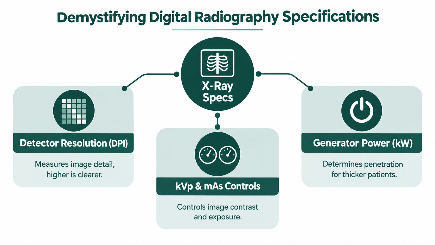
A specification sheet becomes useful when you translate each item into a clinical question. Don't ask whether a number looks impressive. Ask what the generator, tube, detector, and software will let your team do reliably with the species and patient sizes you treat.
Read generator power as a patient-size requirement
Veterinary digital radiography systems commonly use high-frequency generators in the 30–32 kW class, with tube voltage ranges of about 40–125 kV and focal spots around 0.6/1.3 mm, as shown in this digital radiography system specification reference. Those figures aren't a universal purchasing target, but they provide a useful benchmark for evaluating whether a proposed system has enough flexibility for your caseload.
Generator power affects penetration capacity. A small-animal practice may focus on feline thoracic studies, canine limbs, and abdominal work, while a mixed or large-animal practice may need substantially more flexibility through thicker anatomy. The key question is whether the system can produce consistent, diagnostically useful images across the patient range without forcing staff into improvised techniques.
Tube voltage, expressed as kV or kVp, influences beam energy and penetration. A broader range gives the operator more control when moving from thin body parts to thicker regions. mAs, the product of tube current and exposure time, affects the amount of radiation used for the exposure and therefore influences image noise and detail.
Treat focal spot size as a detail and workload decision
A smaller focal spot can support better spatial detail, particularly when the clinical question involves fine bony structures. The larger focal spot setting can support higher loading when the anatomy or workflow demands it. Ask the supplier how operators select between focal spots and whether the system manages those choices clearly during routine use.
Detector resolution matters, but it shouldn't be evaluated in isolation. A high-resolution detector won't rescue poor positioning, motion, inadequate collimation, or an exposure technique that doesn't match the patient. Review sample images from the body regions your team handles most often, and ask who produced them, under what technique, and with what degree of positioning support.
Demand technique discipline
Image quality depends heavily on dose and geometry. A veterinary radiology study found that increasing mAs improved low-contrast resolution and reduced noise, while better collimation, optimized source-to-image distance, and controlled object-to-detector geometry were needed to keep radiographs diagnostically acceptable. The veterinary radiology study on image quality and technique supports a practical conclusion: equipment capability and staff technique must be purchased and managed together.
Require a hands-on demonstration that covers:
- A thin body region where fine detail matters.
- A thicker patient or body region where penetration is the concern.
- A repeat-prone study from your current workflow.
- Image review, annotation, export, and storage.
- Detector handling, cleaning, positioning, and damage protection.
A machine that looks strong in a demonstration can still underperform if your staff find its controls confusing or if the detector doesn't suit the way your rooms operate.
Evaluating True Costs and Market Realities
The quoted equipment price is only one line in the investment. Your budget must include preparation, delivery, installation, integration, training, service, and future replacement exposure. A low initial quote can become the expensive choice if it excludes the work required to make the system usable.
The market is large enough to justify disciplined procurement. The global veterinary x-ray market was estimated at USD 827.5 million in 2025 and is projected to reach USD 1.6 billion by 2035, implying roughly 93% growth over the decade at a 6.9% CAGR, according to this veterinary x-ray market analysis. The same source identifies the equipment segment at USD 352 million in 2025, which indicates that hardware remains the central spending category in this niche.
Build a total-cost view
| Cost Category | Estimated Range | Frequency |
|---|---|---|
| Equipment purchase | Vendor-specific | One-time |
| Room preparation and installation | Vendor and site-specific | One-time |
| Software and image management | Contract-specific | Initial and recurring |
| Integration with practice systems | Integration-specific | Initial, with possible support costs |
| Staff training | Training-plan specific | Initial and refresher |
| Preventive maintenance | Service-contract specific | Recurring |
| Detector repair or replacement reserve | Risk-dependent | Periodic or contingency |
| Compliance and safety requirements | Site-specific | Initial and recurring |
These categories are planning headings, not invented price ranges. Obtain written figures for each line and ask what happens when the included support period ends. Review the total cost of capital considerations before comparing financing offers, because the lowest monthly payment may carry a longer repayment period or additional fees.
Separate essential cost from optional cost
Installation and room readiness are usually essential. Software modules, advanced workflow features, extended warranties, and integration services require a sharper judgment. Pay for features that address a documented clinical or operational need. Don't purchase optional functionality just because it appears on a premium configuration.
Maintenance deserves particular attention. Ask how service is delivered, what parts are covered, how detector damage is handled, and whether software support continues after the initial term. A practice should also reserve for an eventual detector problem even when the manufacturer presents the system as durable.
Finally, model the purchase under conservative utilization. Estimate how the equipment behaves if appointment demand softens, a clinician leaves, or another capital need appears. A sound investment should remain manageable under ordinary business stress, not only under an optimistic growth scenario.
Comparing Acquisition Strategies for Clinics
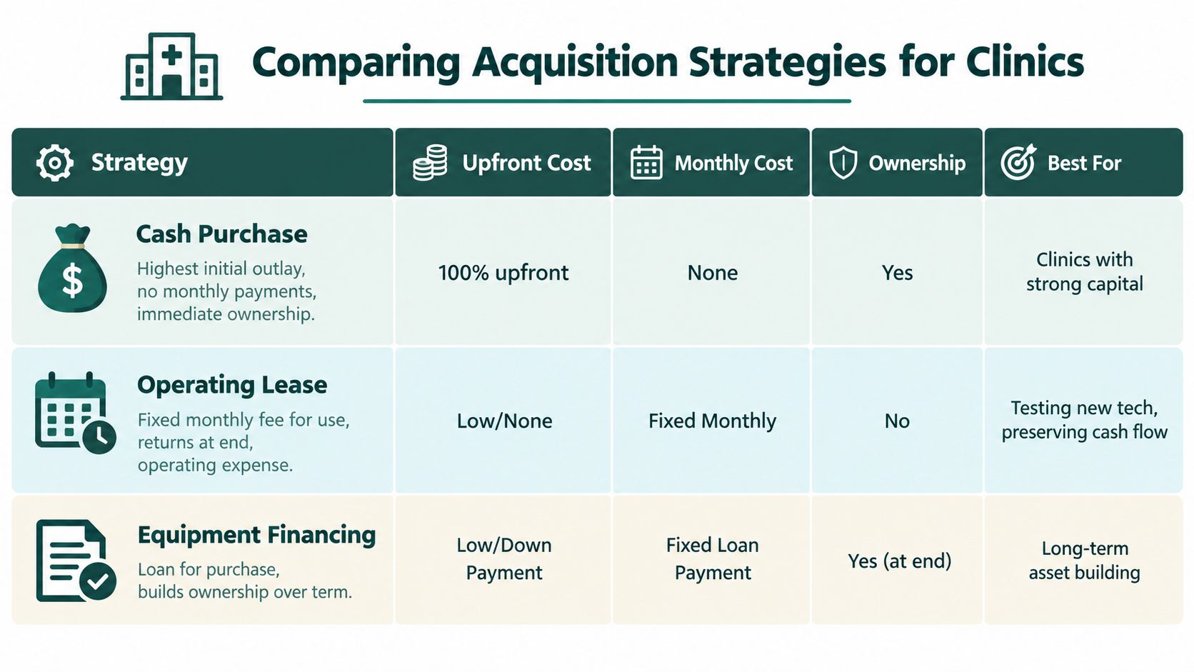
The best acquisition structure depends on your liquidity, tax advice, ownership goals, and confidence in future utilization. There isn't one correct answer for every practice, but there is a wrong answer for each financial situation. A clinic with thin reserves shouldn't empty its operating account just to avoid a loan, and a well-capitalized practice shouldn't borrow automatically without comparing the opportunity cost of its cash.
Cash purchase
Paying cash gives the clinic immediate ownership and avoids scheduled debt payments. It can also simplify the transaction and reduce exposure to lender covenants. The trade-off is liquidity. Once the funds leave the business, they can't cover a payroll spike, urgent facility work, inventory needs, or another opportunity.
Cash can make sense when reserves remain strong after the purchase and when the practice has no more productive use for the capital. Don't confuse a debt-free asset with a financially healthy practice. A paid-for machine can still create pressure if the clinic has insufficient working capital around it.
Operating lease
An operating lease generally provides access for a fixed payment without creating the same ownership position as a purchase. This can suit a clinic that wants flexibility, expects technology preferences to change, or wants to preserve cash for expansion.
The central disadvantage is residual value. Depending on the contract, the practice may return the equipment without building equity. Read the end-of-term provisions, renewal language, purchase option, maintenance responsibility, and consequences of early termination. A lease isn't automatically cheaper because its initial cash requirement is lower.
Equipment financing
Equipment financing funds the purchase while allowing the clinic to preserve more cash than an outright purchase. The practice makes scheduled payments and generally builds ownership in the asset over the repayment period. This structure often fits an established clinic that expects to use the system for years and wants the asset to remain on its balance sheet.
Tax treatment depends on the agreement and applicable rules, so involve your accountant before signing. Ask how depreciation, interest, ownership, and end-of-term obligations will be treated. The equipment financing and leasing guidance can help frame the questions, but the final structure should reflect your practice's financial statements and tax advice.
| Strategy | Liquidity effect | Ownership position | Strongest fit |
|---|---|---|---|
| Cash purchase | Highest immediate cash reduction | Immediate ownership | Strong reserves and predictable needs |
| Operating lease | Preserves cash through scheduled use payments | Usually no ownership at return | Flexibility and technology testing |
| Equipment financing | Preserves more working capital than cash | Ownership develops through repayment | Long-term use and asset building |
The right structure leaves the clinic able to operate well after the equipment arrives.
Aligning Loan Amortization with Equipment Lifecycle
Set the financing term against the system's dependable useful life, not the lowest available monthly payment. A longer term can ease near-term cash pressure, yet it may leave the clinic paying for equipment after its clinical value, software support, or integration has declined.
Imaging equipment ages as a system, but its components do not age evenly. The generator, tube, detector, workstation, and image-management environment carry different replacement and support risks. Build the repayment plan around the full operating system rather than the equipment line on the invoice.
Match repayment to dependable utilization
Begin with revenue and costs you can defend. Identify the studies the system will support, staff time it will save, referrals it may retain, and additional operating costs it will create. Model the expected cash contribution under lower demand and seasonal variation before accepting loan terms.
The monthly payment should be covered by durable operating capacity, not a best-case forecast. If the equipment must run at exceptionally high utilization to meet the obligation, the structure is too aggressive. If repayment extends beyond the point when the practice expects to replace the system, the term is too long.
Use a replacement reserve alongside the loan. Allocate room for maintenance, training, software-related requirements, and future equipment purchases. The clinic should not have to choose between servicing the current system and preparing for its replacement.
Consider obsolescence separately from physical durability
A machine can remain functional while becoming harder to support or less useful in daily workflow. Integration requirements, detector performance expectations, staff training, and image-management needs can change before a major component fails. Market history provides context, but it should not justify assuming that every current system will retain the same useful life.
Ask the supplier and lender:
- Which components are most likely to require replacement?
- How long will software and integration support remain available?
- Can the repayment schedule accommodate a planned upgrade?
- What happens if the practice sells, relocates, or adds another site?
- Are there prepayment restrictions or collateral requirements?
Document the expected replacement window before signing. Then compare that window with the final payment date. A sound structure lets the asset generate clinical and financial value while it remains relevant, with enough cash-flow capacity left for reinvestment.
Securing Veterinary Focused Equipment Financing
A strong application explains how the equipment fits the clinic's operations. Lenders need more than a quote. They need to understand the practice's revenue pattern, existing obligations, cash reserves, and the specific role the system will play in patient care and workflow.
Prepare the financial record
Gather complete financial materials before requesting terms:
- Historical statements: Provide profit and loss statements, balance sheets, business tax returns, and current debt information.
- Banking evidence: Organize deposits and operating-account activity so the lender can see how revenue moves through the practice.
- Equipment proposal: Include the vendor quote, installation scope, software, service coverage, training, and expected delivery schedule.
- Operating forecast: Show how the equipment fits expected caseload, staffing, pricing, referral retention, and expenses.
- Ownership information: Clarify the legal entity, ownership shares, guarantors, and any planned expansion.
The forecast shouldn't promise dramatic gains without support. Use your own appointment data, referral patterns, current imaging volume, and documented bottlenecks. A conservative model often earns more credibility than an aggressive projection that depends on perfect scheduling.
Explain clinical necessity in business terms
Describe which studies the current system handles poorly, which cases leave the practice, and where staff lose time. Then connect the proposed system to capacity, service continuity, and diagnostic confidence. The lender doesn't need a radiology lecture, but the underwriter does need to see why the purchase belongs in the clinic's capital plan.
A veterinary-focused lender may evaluate clinic deposits, cash flow, and operating patterns alongside credit history. That matters because veterinary revenue can be shaped by appointment cycles, pharmacy purchases, emergency demand, staffing changes, and seasonal fluctuations. Present those realities clearly rather than forcing the practice into a generic small-business narrative.
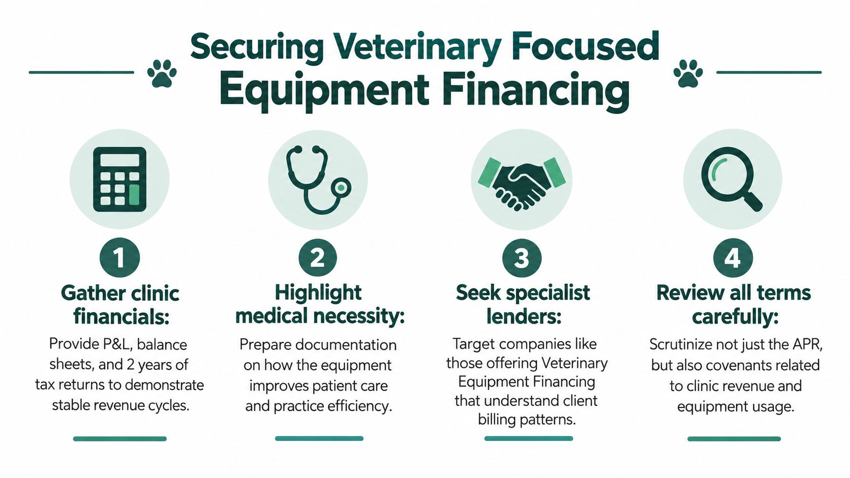
Before signing, compare total repayment, fees, collateral, personal guarantees, insurance requirements, prepayment terms, and any financial covenants. A fast approval isn't useful if the structure limits future working capital or creates pressure during slower periods.
Use the following video as a prompt for your internal discussion, then verify every financing term against the written agreement and your professional advisers.
Maximizing Diagnostic Quality and Return on Investment
The machine won't produce a return by itself. Your team creates that return through consistent positioning, exposure selection, detector care, image review, and scheduling discipline. The veterinary radiology evidence cited earlier makes the operational point clear. Dose, collimation, distance, and geometry all influence whether the image answers the clinical question.
Create a short implementation plan before installation:
- Assign ownership: Name the clinical lead responsible for protocols and the operational lead responsible for uptime and supplies.
- Standardize common studies: Build technique guidance for the body regions your practice handles most often.
- Train by workflow: Teach positioning, detector handling, image review, retakes, cleaning, and escalation procedures.
- Audit retakes: Review repeated studies for preventable causes rather than blaming individual staff members.
- Track utilization: Monitor how often the system supports in-house care, referral retention, and urgent decision-making.
Training should include the people who position patients, operate the system, review images, and manage records. A technically capable machine can underperform when only one experienced employee knows how to use it. Cross-training also reduces operational risk when that employee is absent.
A radiography asset earns its place through repeatable use, not occasional impressive images.
Keep financial and clinical reviews connected. If utilization falls, find out whether the problem is demand, scheduling, confidence, positioning, maintenance, or referral habits. If retakes rise, investigate the protocol and training before assuming the equipment is defective. If the clinic is considering related imaging, evaluate the broader capital plan through the same discipline used for veterinary ultrasound equipment, including utilization, lifecycle, integration, and cash-flow impact.
The strongest purchase is one that improves patient care without forcing the practice to sacrifice resilience. Choose specifications that match the caseload, price the complete lifecycle, protect working capital, and align repayment with useful service. Then give the team the training and accountability needed to turn the system into a dependable clinical and financial asset.
Veterinary Practice Loans offers veterinary-focused equipment financing and leasing structures designed around clinic cash flow, asset useful life, and long-term ownership goals. Visit Veterinary Practice Loans to discuss your veterinary x ray equipment purchase, compare acquisition options, and build a funding plan before you commit to a quote.
